A high-precision ocular biometry platform for advanced glaucoma diagnostics and surgical planning.
New York Eye and Ear Infirmary of Mount Sinai
Jun 2022 – Present
Glaucoma surgery is done to help fluid drain out of the eye, but until now nobody could measure that drainage during the operation. Surgeons adjust drainage devices by feel and find out days later whether the eye drains too much or too little. Too much drainage drops the pressure too low (hypotony), which can cause the eye to collapse, bleed or lose vision.
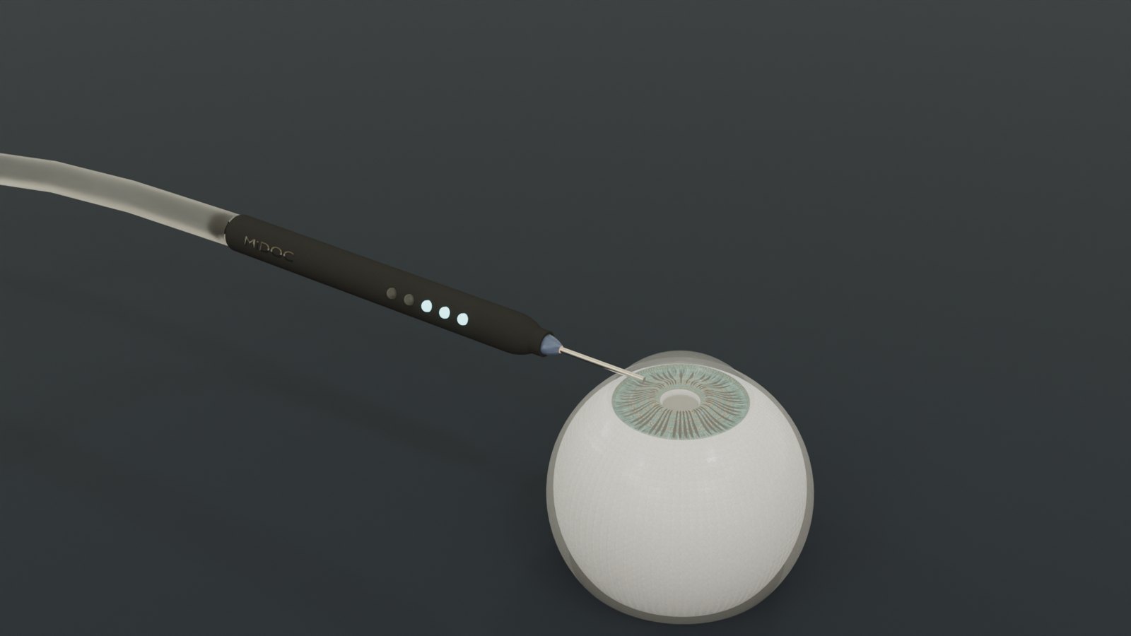
miDOC measures drainage in the operating room. I helped invent it, built the research system, ran a first-in-human feasibility study, and now lead the engineering of the commercial version.
Through a small cannula in a standard side incision, miDOC holds the eye at a set pressure and measures how much fluid it takes to keep it there. At steady state, what goes in is what drains out. Doing that at two pressures gives the eye's outflow facility, the extra drainage per mmHg:
C = ΔQ / ΔP
Measuring at the start and end of surgery shows what the operation actually changed. Older methods (tonography, pressure decay) only estimate drainage and depend on assumptions about how stiff the eye is. Holding pressure constant and measuring flow directly removes that assumption.
In a first-in-human study at New York Eye and Ear Infirmary of Mount Sinai, the change in flow during surgery flagged every measured patient who later developed early hypotony, with no false alarms. The device was introduced at ASCRS in April 2026. The manuscript is in preparation, so the detailed numbers will go here once it's published.
On the bench, the system was validated against a precision syringe pump and a set of glass capillaries with known drainage, including a simulated heartbeat.
I co-invented miDOC and its workflow. I designed and built the research system (mechanics, fluidics, electronics, firmware and analysis), ran the bench validation and the surgical cases, co-wrote the first-in-human manuscript, and designed the commercial version for mass manufacture and FDA compliance.
More from this category.